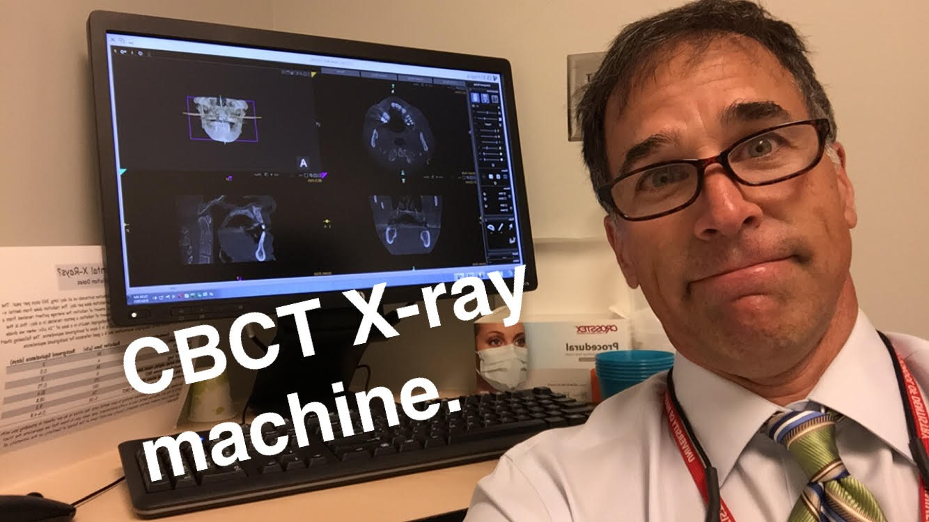
CBCT systems used by dental professionals rotate around the patient, capturing data using a cone-shaped X-ray beam. This data is used to reconstruct a three-dimensional (3D) image of the of the patient’s head and neck. CBCT uses more radiation than regular dental teeth x-rays, but still less than 10% of the radiation used in conventional medical CT scan of the same area.
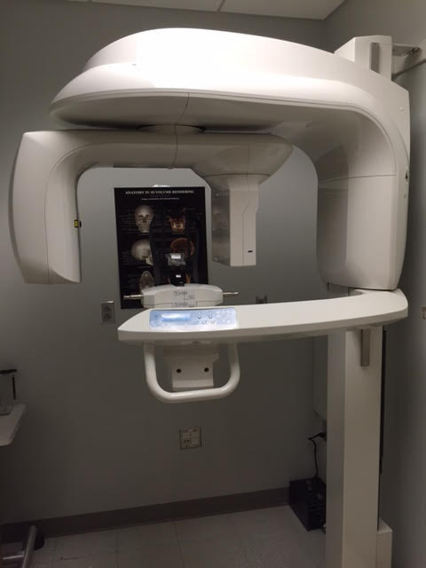
This is the Carestream 9300 Imaging machine we have at the University of Maryland Dental School. I mostly use this to plan dental implant placement.
Dentists use Cone Beam CT imaging for the following:• 3-D observation of overall oral/facial bony characteristics, allowing easier diagnosis and placement of dental implants • Surgical guide fabrication for implant placement • 3-D observation of teeth for endodontic diagnosis and treatment • Diagnosis and treatment of tooth impactions • Identification of inferior alveolar nerve and mental foramen location • Identification of the location of the maxillary sinus • Identification of the presence of odontogenic lesions • Trauma evaluation and treatment • Analysis of temporomandibular joint characteristics leading to diagnosis and treatment • Integration with CAD/CAM devices for fabrication of prosthodontics or orthodontic appliances • Identification for referral of numerous conditions or diseases not normally within the realm of dentistry, but that can be shown on typical cone beam images.


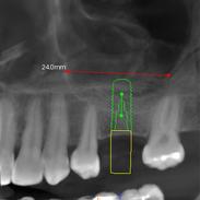
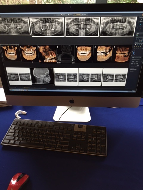
These last 2 pictures show how we use information obtained from the Cone Beam CT to plan placement of dental implants.