For 44 years, I have been practicing dental hygiene in Virginia. Recently I had to have a crown prep done on #8 and a replacement crown on #9. My first crown on #9 was stainless steel, placed at age 7 in 1958. Since then this tooth has had 14 replacement crowns that improved the appearance as dental materials progressed over the years.
My new ones though are the best I’ve ever had made. The contacts, size, shape, color & occlusion are perfect and needed no adjustments when they were delivered. I experienced no discomfort during the procedure, even while wearing the temporaries.
I have seen thousands of crowns & fillings by hundreds of dentists over my long career and Dr. Gentry’s work surpasses them all. He is extremely attentive to detail. The perfect fit and aesthetics are evident in every restoration he does. I have no reservations in recommending his work to family, friends & current patients. His sensitivity and approach while interacting with patients is a model for all individuals interested in entering the profession.
I truly feel he is deserving of the recognition equal to his daily accomplishments in the field of dentistry. As a dental hygienist with 44 years of private practice experience, I know who the best dentists are, and Dr. Gentry is one of the best!
I highly recommend Dr. Gentry. His dental work is the best I have seen.
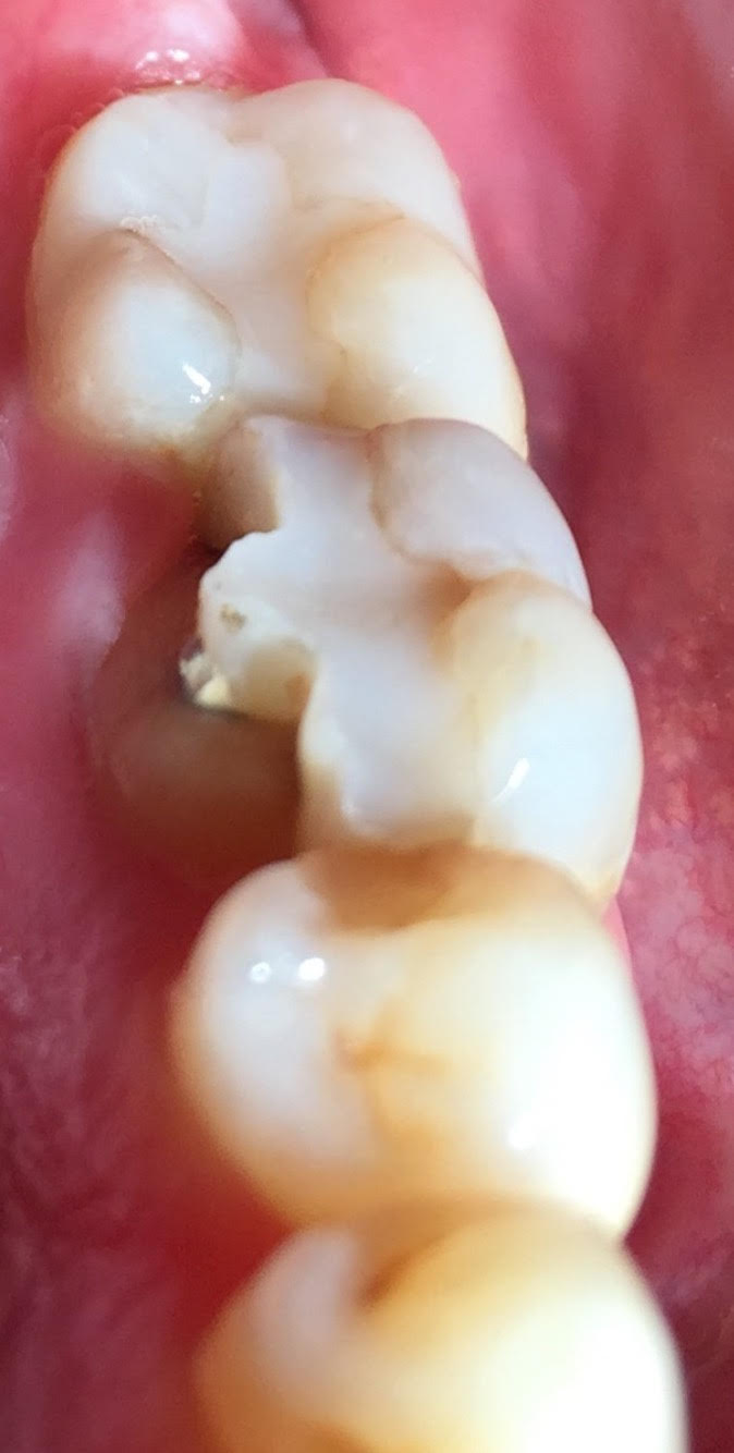
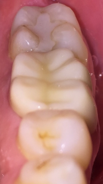
This patient fractured the lingual side of his first molar tooth biting down on a Skittle candy. Dr. Gentry was able to restore the tooth with a porcelain crown and was able to avoid a root canal and crown lengthening periodontal surgery.
Fractured molar tooth.Tooth immediately following crown placement.
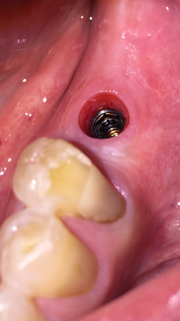
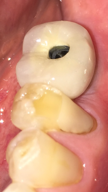
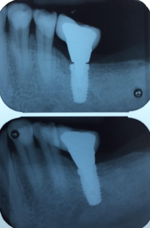
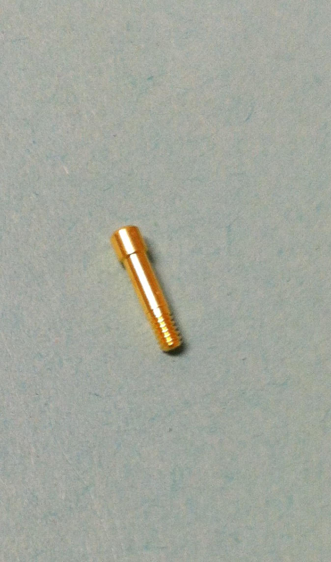
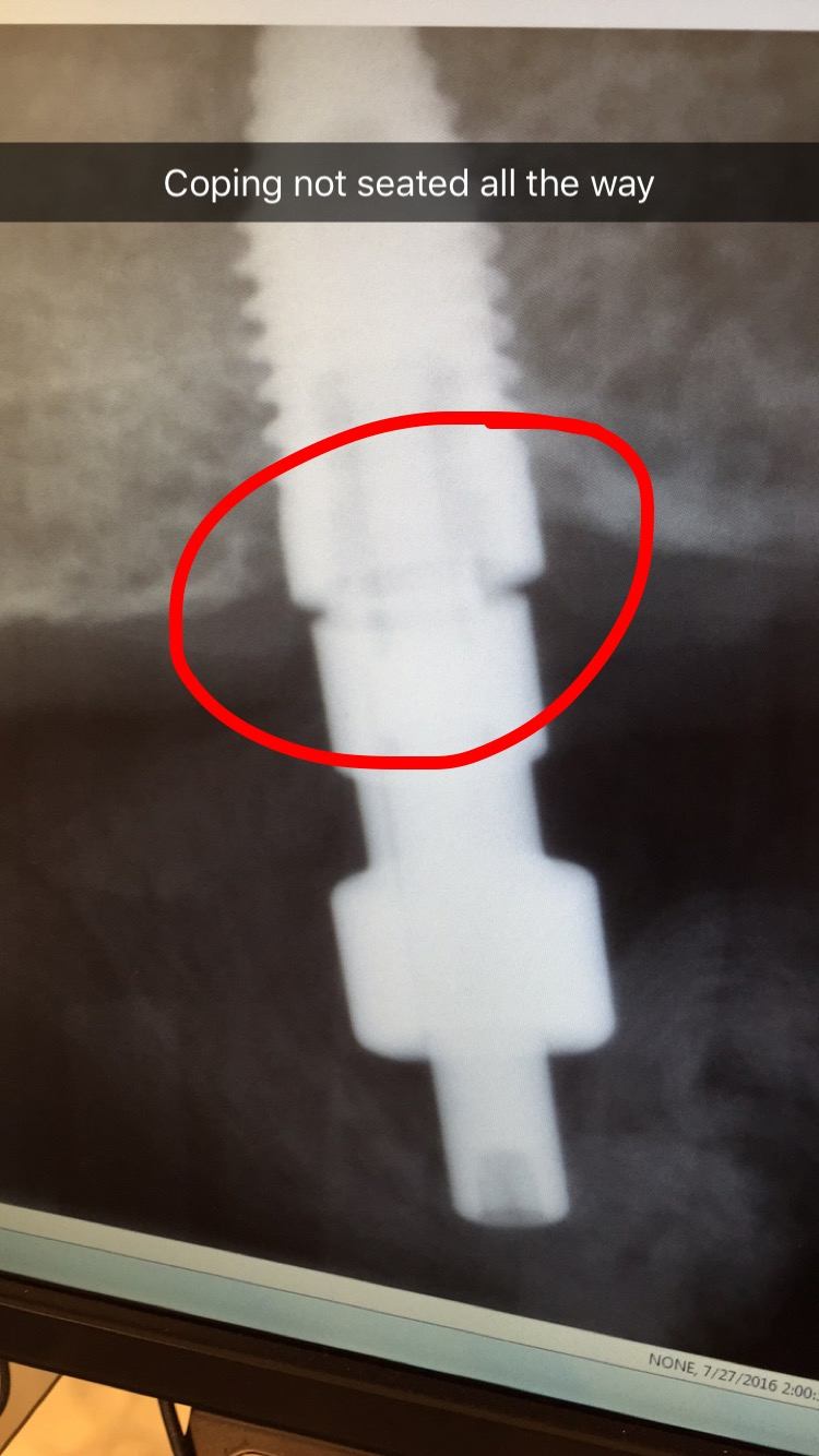
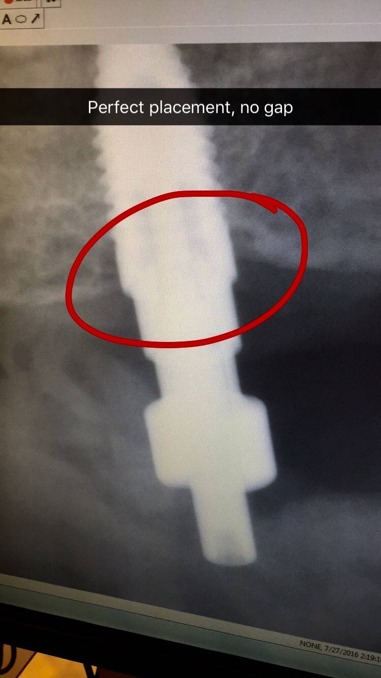
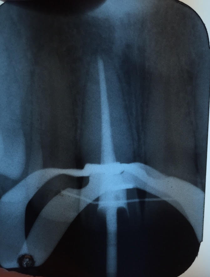
In this case report Dr. Gentry demonstrates the importance of taking a radiograph when seating the custom abutment and crown before final torquing of the gold implant interface connecting screw.
ImplantCustom abutment and crown placed with gold screw tightened to finger pressure.Upper radiograph shows improper interface-note the gap present at the interface. Lower radiograph shows perfect fit, taken after adjusting the contact and re-positioning the crown/abutment on the implant.Gold screw used to attach crown and abutment to implant.Now the gold screw can be tightened to 20 Newtons per centimeter squared with the torque wrench.Screw hole filled in with composite filling material.
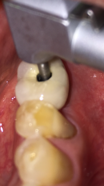
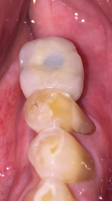
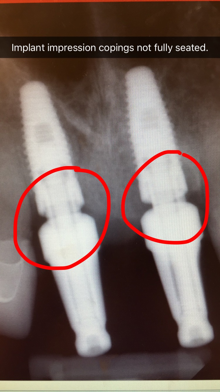
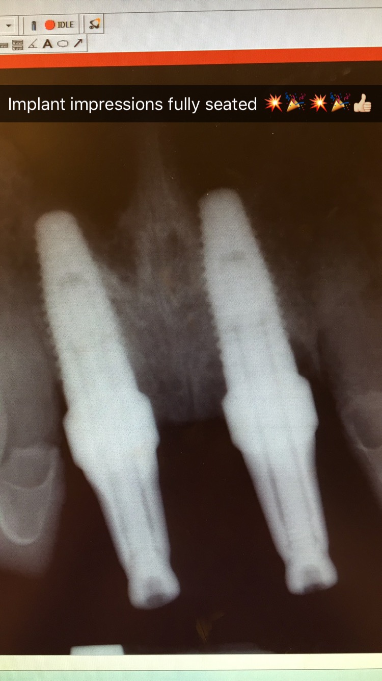
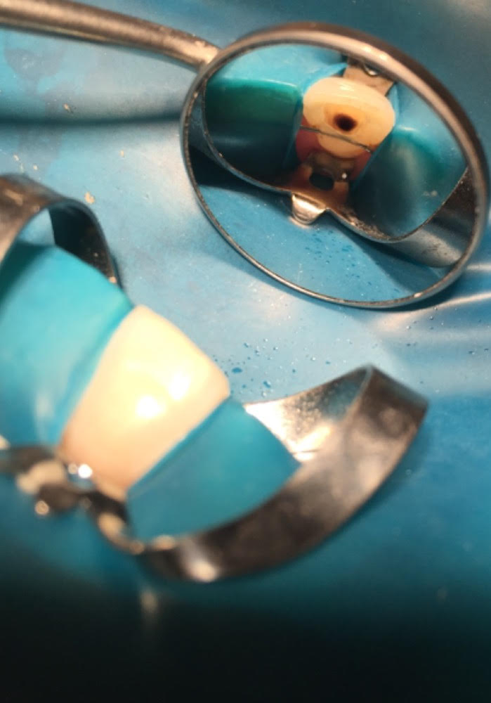
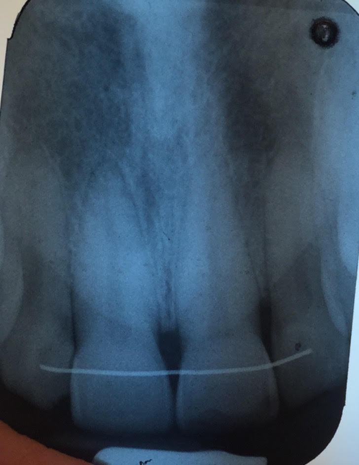
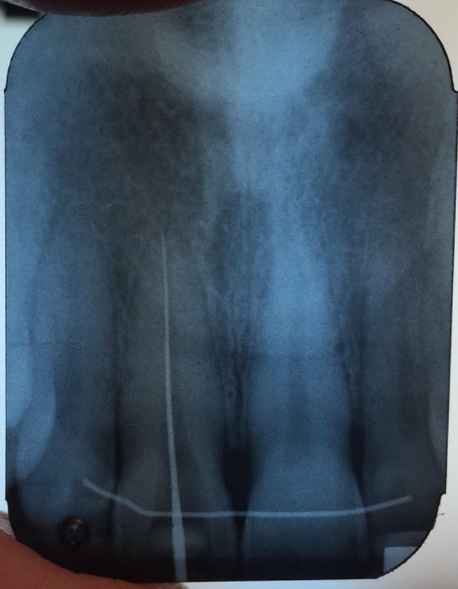
Here’s an example from a case I was doing with a dental student involving the 2 upper front teeth. In the first x-ray the impression copings were not fully seated. I re-positioned the copings and another x-ray was taken to confirm proper alignment. You cannot tell without an x-ray since this is below the gum-line.
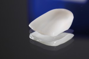
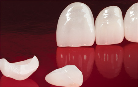
Porcelain veneers are thin pieces of porcelain used to recreate the natural look of teeth, while also providing strength and resilience comparable to natural tooth enamel. It is often the material of choice for those looking to make slight position alterations, or to change tooth shape, size, and/or color.
VENEER CONSULTATION
The first step is to schedule an appointment with Dr. Gentry to determine if veneers are the right option for you, or if there are alternate solutions available. Communication about what you want corrected is critical for a successful result. Spend time clearly identifying what cosmetic improvements you want to accomplish.
You’ll often hear people say that celebrities have veneers and this may seem like the best way to replicate picture-perfect teeth, but each mouth is different and veneers need to be carefully researched. We begin with a smile analysis and diagnostic wax mock-up that will allow you to see if the final result is actually what you’re looking for. Deciding that porcelain veneers will create the look you want is only one step in the process. There is much more to learn before proceeding further.
THE HOWS AND WHYS OF PORCELAIN VENEERS
Porcelain laminate veneers consist of a compilation of several thin ceramic layers which replace original tooth enamel, and an adhesive layer. To apply a veneer, a very small amount of the original tooth enamel must be removed, usually about a millimeter. This is essential as it creates room for the porcelain veneer to fit within the mouth and most accurately restore natural tooth function while creating an even better appearance than the original tooth.
The bond between original tooth and porcelain veneer is critical as it not only provides the esthetic perfection desired, but also a strong bond which is essential for correct veneer function. Light-sensitive resin is placed between the original tooth and the veneer and then hardened using a special curing light.
Porcelain veneers are a very successful option in many situations where the original tooth has developed poor color, shape, and contours. It is also a good choice for fractured teeth, gaps between teeth, and in some situations where the tooth position is compromised and there are minor bite-related problems. For some people, superficial stains do not respond well to tooth whitening or bleaching. In these situations, a porcelain veneer may be the best option.
THE BENEFITS OF VENEERS
Since veneers are individually sculpted for each patient, it is nearly impossible to tell the difference between a veneer and a natural tooth. Unlike natural teeth, custom-made veneers resist coffee and tea stains, and cigarette smoke because they are made of high-tech materials.
With veneers—as opposed to crowns—your natural teeth remain largely intact with only a minimal amount being altered to fit the veneer.
For teeth that resist whitening, veneers can make even the darkest teeth appear bright white.
Dentists may also recommend veneers to quickly fix minor twists, overlaps, and small gaps.
POTENTIAL VENEER DOWNSIDES
Because a portion of the original tooth enamel is reduced, a veneer is not considered a reversible treatment. Although adjustments and even new veneers can be made, you can never reliably return to the original condition of the tooth.
Creating porcelain veneers requires some laboratory time, so expect at least a week before they’re ready to be applied.
After the porcelain veneers are attached you will probably have some sensitivity to hot and cold temperatures due to the removal of that thin layer of enamel. This typically disappears within a few days. In a healthy mouth properly treated with porcelain veneers—and where destructive forces are minimized or eliminated—a patient should be able to use porcelain veneers like his or her own teeth. Although they’re very strong, veneers are also brittle. You should avoid the same excessive stresses you would avoid with non-veneered teeth: don’t bite your fingernails, chew ice, or open beer bottles with your veneers!
MAINTENANCE OF A PORCELAIN VENEER
Maintaining porcelain veneers is actually quite simple: Treat them as you would your original teeth, with routine brushing and flossing. Using non-abrasive fluoride toothpaste will typically be suggested by your dental professional.
One week after your veneers are placed, you will be required to return to the office for a follow-up visit and evaluation so the dentist can see how your mouth is reacting to the veneers. Even if you feel the veneers are a success, this appointment is vital to your future oral health.
If you have a habit of grinding or clenching your teeth, we will make you a nighttime bite guard so you do not damage your veneers.
You should also return for regular professional maintenance because porcelain veneers should be polished with a specially formulated, non-abrasive paste, and because we need to inspect your dentistry for any sign of potential failure.
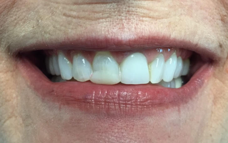
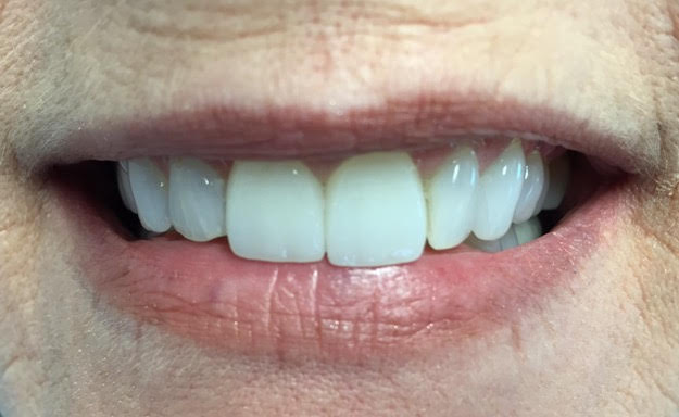
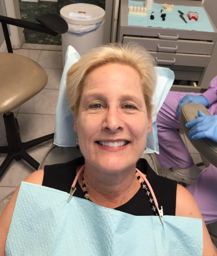
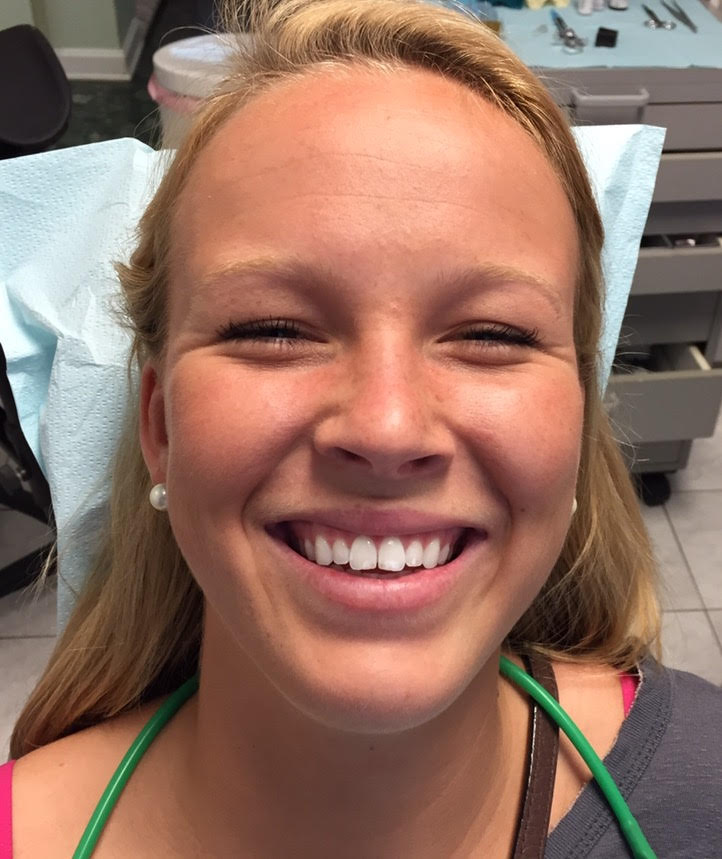
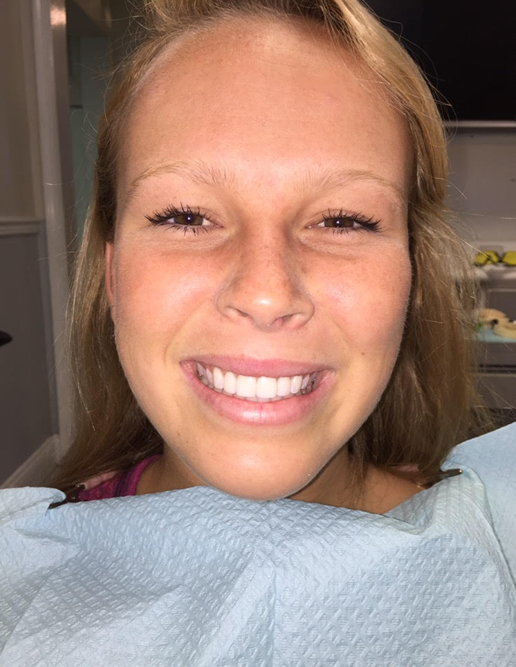
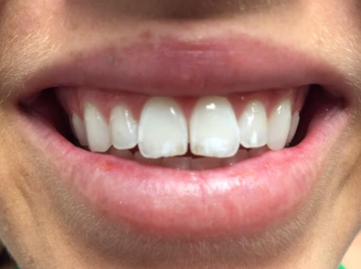
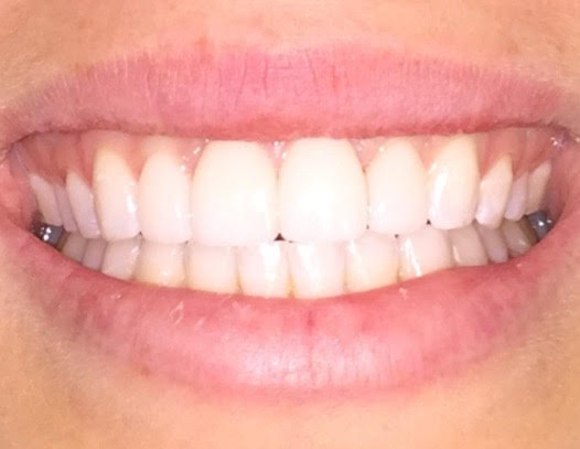
One of Dr. Gentry’s Veneer Patients from last week
BeforeAfter. She is numb so it’s difficult to smile.BeforeAfter
To understand root canal treatment, it helps to know something about the anatomy of the tooth. Inside the tooth, under the white enamel and a hard layer called the dentin, is a soft tissue called the pulp. The pulp contains blood vessels, nerves, and connective tissue and creates the surrounding hard tissues of the tooth during development.
The pulp extends from the crown of the tooth to the tip of the roots where it connects to the tissues surrounding the root. The pulp is important during a tooth’s growth and development. However, once a tooth is fully mature it can survive without the pulp, because the tooth continues to be nourished by the tissues surrounding it
Why would I need a root canal?
A root canal is necessary when the pulp, the soft tissue inside the root canal, becomes inflamed or infected. The inflammation or infection can have a variety of causes: deep decay, or a crack or chip in the tooth. In addition, an injury to a tooth may cause pulp damage even if the tooth has no visible chips or cracks. If pulp inflammation or infection is left untreated, it can cause pain or lead to an abscess.
What are the signs of needing root canal treatment?
Signs to look for include pain, prolonged sensitivity to heat or cold, tenderness to touch and chewing, discoloration of the tooth, and swelling, drainage and tenderness in the lymph nodes as well as nearby bone and gum tissues. Sometimes, however, there are no symptoms.
How does root canal treatment save the tooth?
The dentist removes the inflamed or infected pulp, carefully cleans and shapes the inside of the root canal, then fills and seals the space. Afterwards, a crown or other restoration on the tooth to protect and restore it to full function. After restoration, the tooth continues to function like any other tooth.
Will I feel pain during or after the procedure?
Many root canal procedures are performed to relieve the pain of toothaches caused by pulp inflammation or infection. With modern techniques and anesthetics, most patients report that they are comfortable during the procedure.
For the first few days after treatment, your tooth may feel sensitive, especially if there was pain or infection before the procedure. This discomfort can be relieved with over-the-counter or prescription medications. Your tooth may continue to feel slightly different from your other teeth for some time after your root canal treatment is completed.
Step-by-Step Root Canal Procedure
Root canal treatment can often be performed in one or two visits and involves the following steps:
1. The dentist examines and x-rays the tooth, then administers local anesthetic. After the tooth is numb, a small protective sheet called a “dental dam” is placed over the area to isolate the tooth and keep it clean and free of saliva during the procedure.
2. An opening in the crown of the tooth. Very small instruments are used to clean the pulp from the pulp chamber and root canals and to shape the space for filling.
3. After the space is cleaned and shaped, the dentist fills the root canals with a biocompatible material, usually a rubber-like material called gutta-percha. The gutta-percha is placed with an adhesive cement to ensure complete sealing of the root canals. In most cases, a temporary filling is placed to close the opening. The temporary filling will be removed usually 1 week later.
Can all teeth be saved with a root canal?
Most teeth can be saved. Occasionally, a tooth can’t be saved because the root canals are not accessible, the root is severely fractured, the tooth doesn’t have adequate bone support, or the tooth cannot be restored.
Here’s photos of what an actual root canal looks like on a patient.
Access opening made by Dr. Gentry on inside surface of tooth.Isolation dam placed over tooth.Before treatment x-ray.Cleaning the root canal.Final root canal fill.
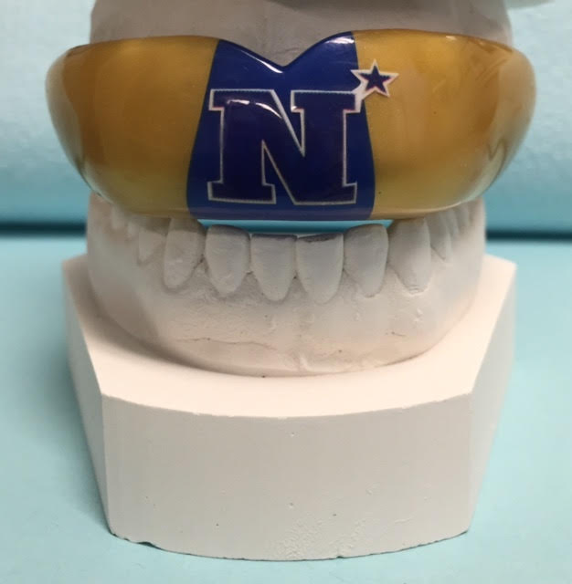
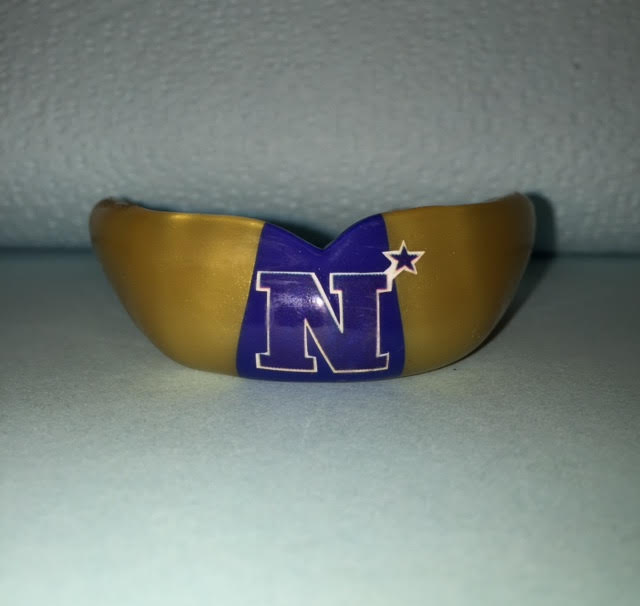
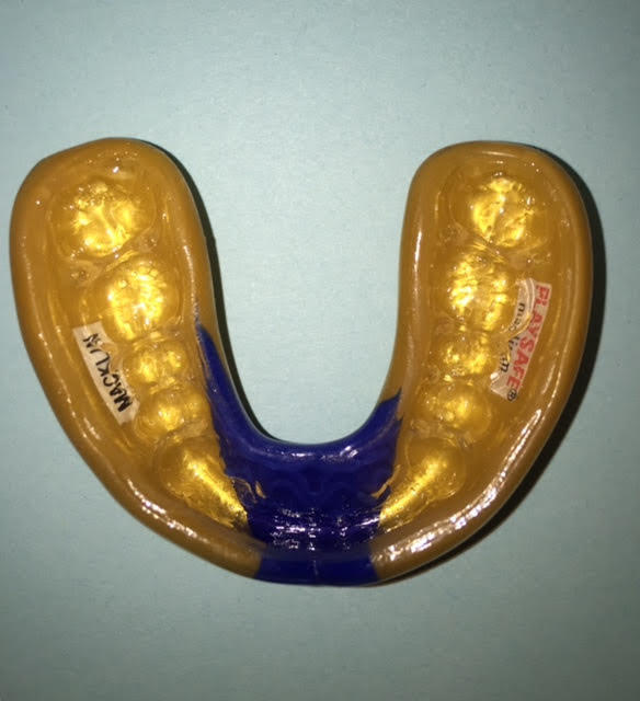
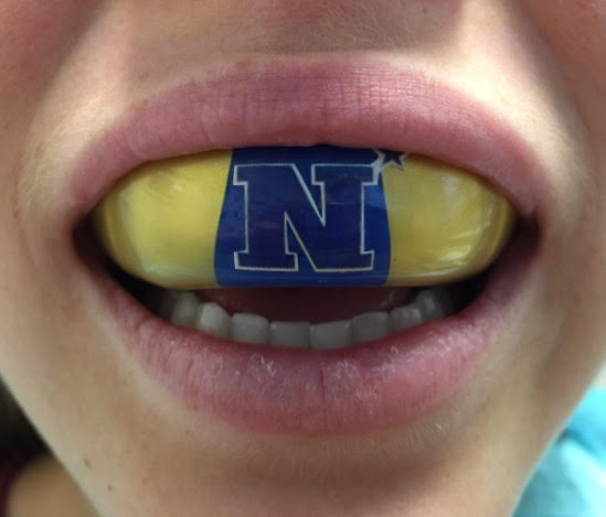
Custom dentist made pressure laminated mouthguards provide the most comfortable fit and the best protection against injury. They are more durable and longer lasting than over the counter mouthguards. I can make the mouthguards in your team colors and with the team logo.
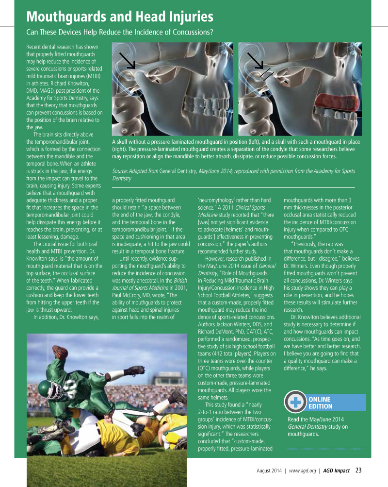
Mouthguards protect your teeth, mouth, face, lips and jaws. Research has shown that custom made mouthguards reduce the incidence of concussions and brain injuries. I have attached below an article from The Academy of General Dentistry Magazine, August 2014, on mouthguards reducing brain injuries.
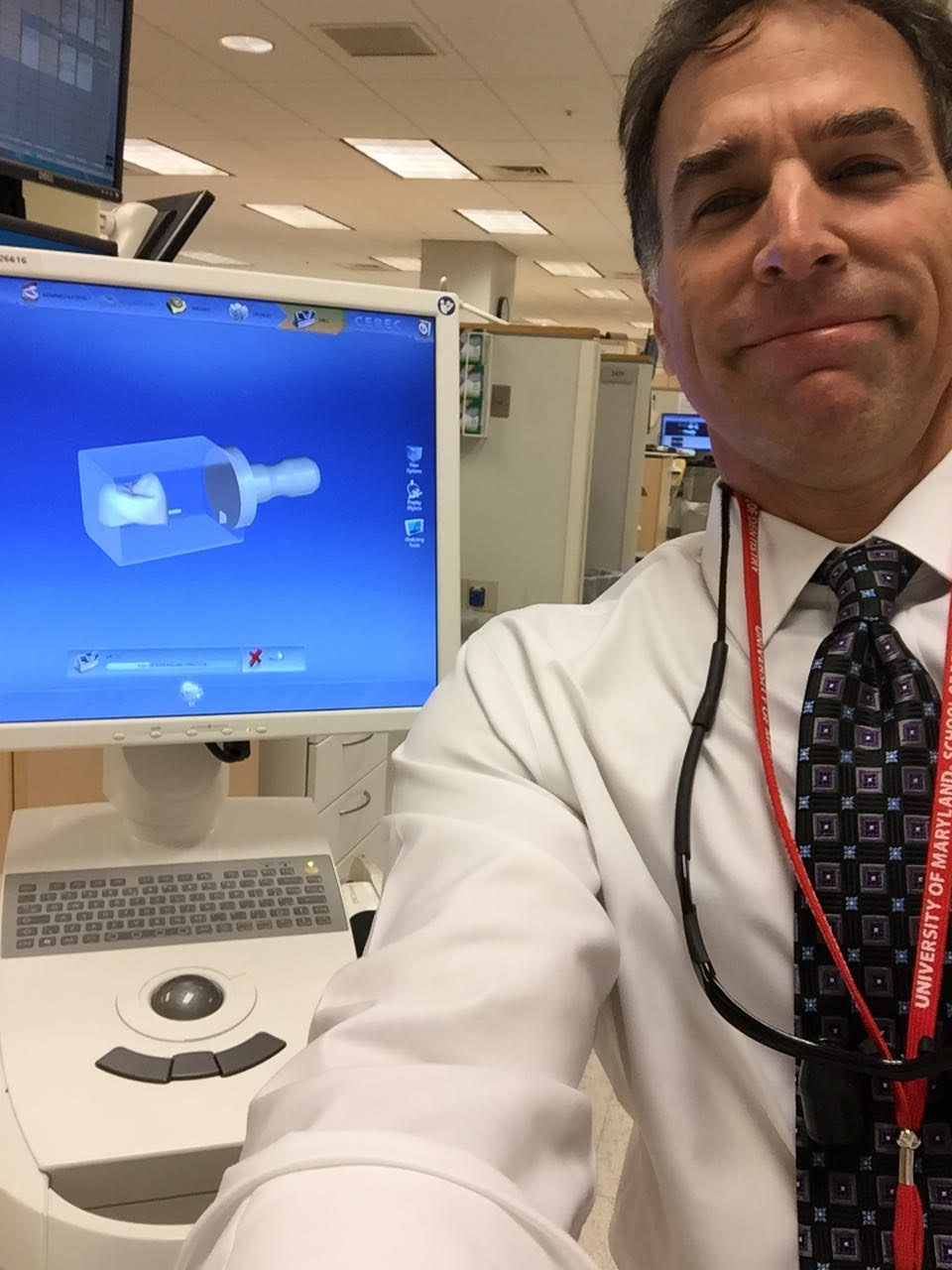
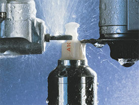
CAD/CAM is an acronym for computer-aided design/computer-aided manufacturing. CAD/CAM technology has been increasingly incorporated into dentistry over the past 20 years. CAD/CAM technology is used by dentists and dental laboratories to provide our patients with 3-D milled ceramic crowns and other types of restorations, and to fabricate abutments for dental implants, used to replace missing teeth. A digital impression is taken of the patient’s teeth or dental impression, and the restoration is milled from a single block of tooth colored ceramic material in a milling chamber.
Digital design of the crown restoration.Sending the digital impression to the 3-D milling machine3-D printer fabricating the dental restoration.
The 3-D milling machine fabricating the dental restoration.