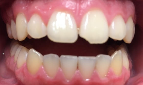
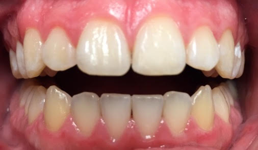
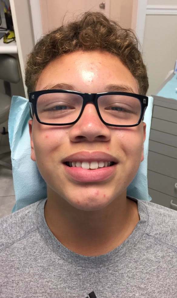
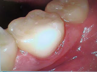
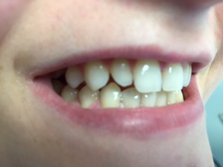
This 14 year old patient chipped his front tooth on the metal restraint bar on a ride at Disney World. Today I repaired the tooth with a ceramic composite bonded restoration in 30 minutes.
This 14 year old patient chipped his front tooth on the metal restraint bar on a ride at Disney World. Today I repaired the tooth with a ceramic composite bonded restoration in 30 minutes.
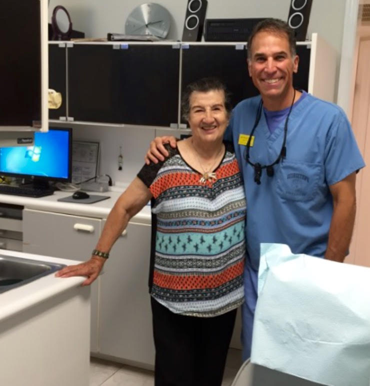
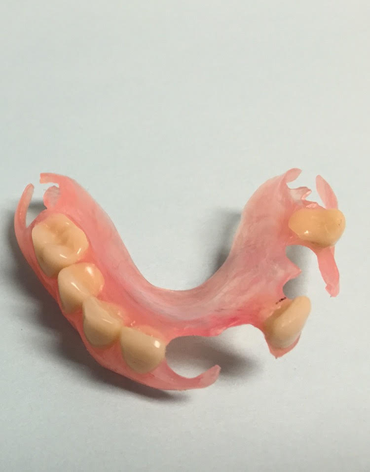
I restored her smile with a Valplast upper partial denture. Nice case :-)))


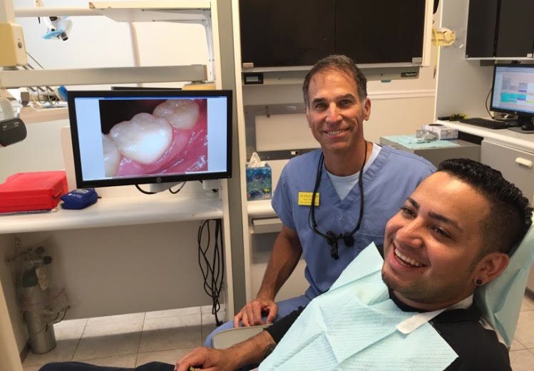
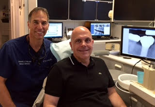
As Dean’s Faculty, Clinical Assistant Professor in The Advanced General Dentistry Department at The University of Maryland School of Dentistry, I am used to teaching and educating residents and senior dental students. I am always happy to answer questions and show patients exactly what I am doing and explain why.
This patient bit down on a fork and fractured his upper left lateral incisor in half. We cleaned and polished his teeth, and I restored the tooth with a porcelain crown. He was very happy.
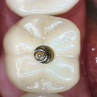
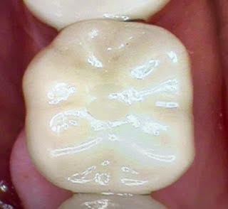
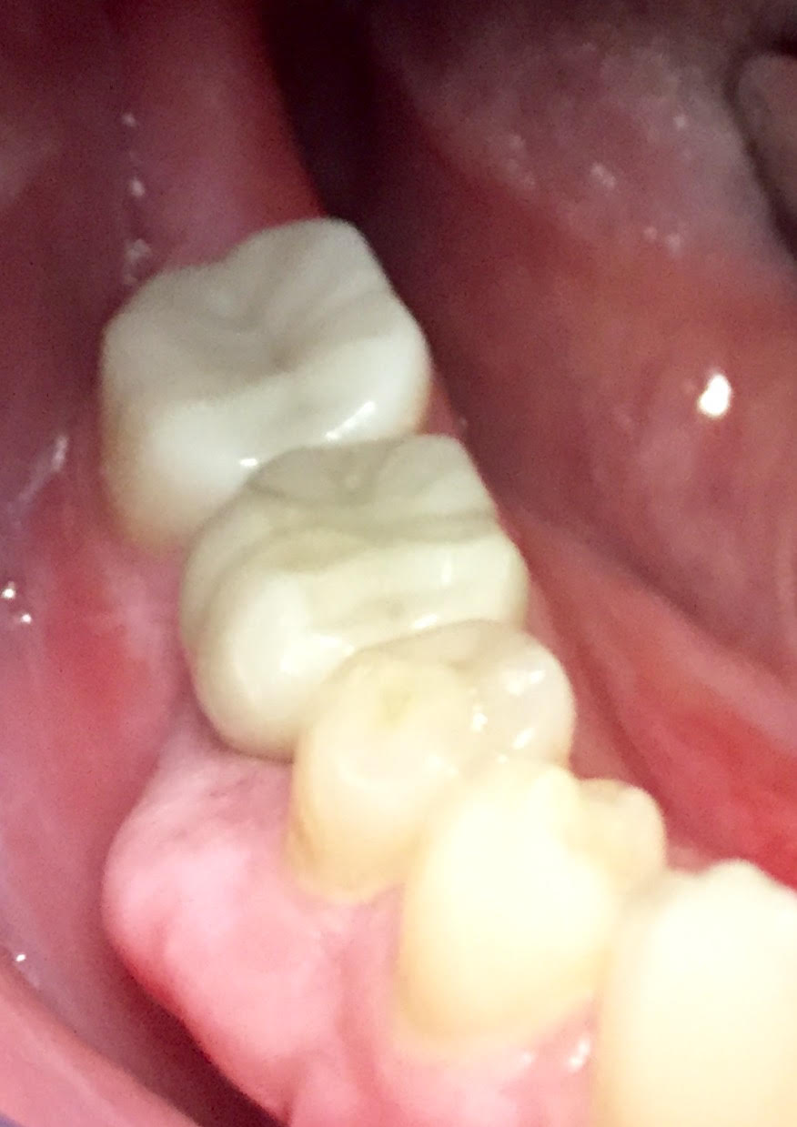
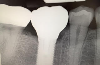
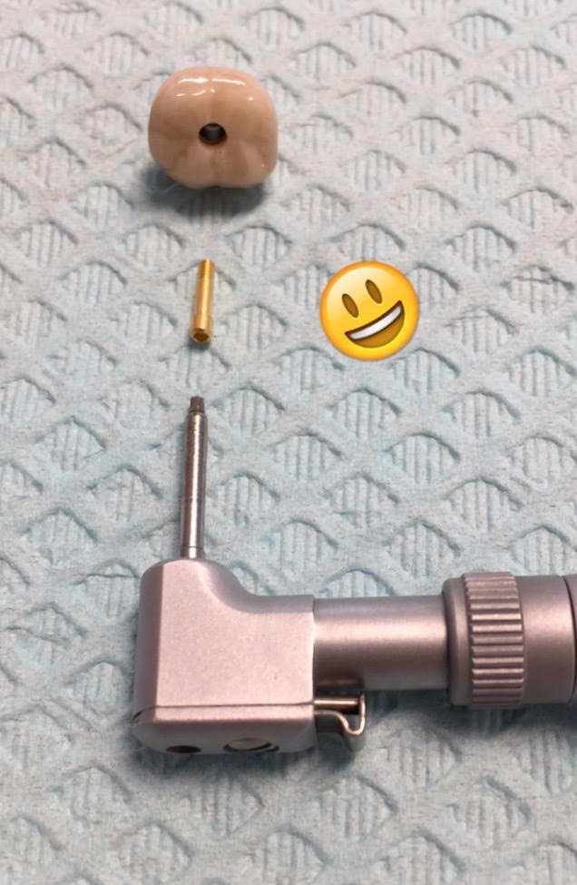
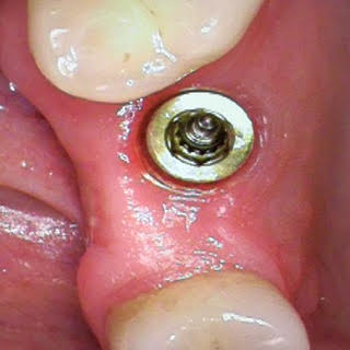
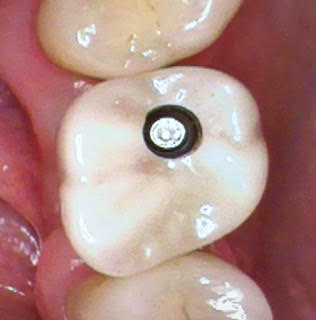
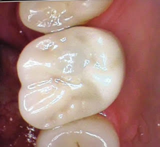
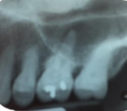
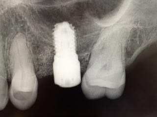
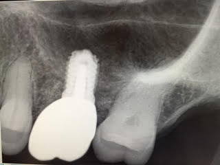
This patient fractured his lower right first molar. We extracted the tooth and placed a 6mm wide 13 mm long Zimmer Biomet Encode titanium implant. This morning I delivered the beautiful porcelain implant crown.
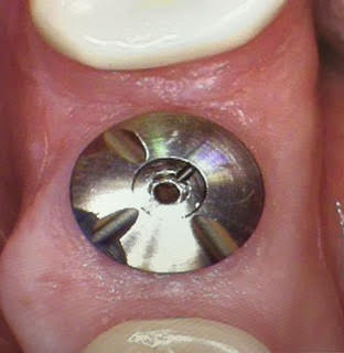
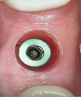
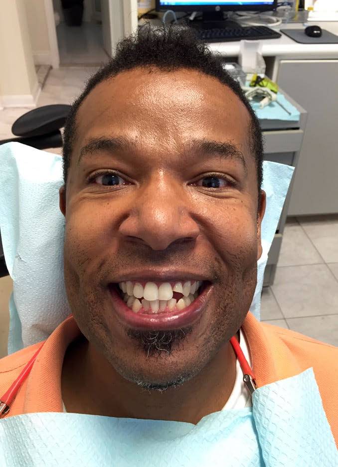
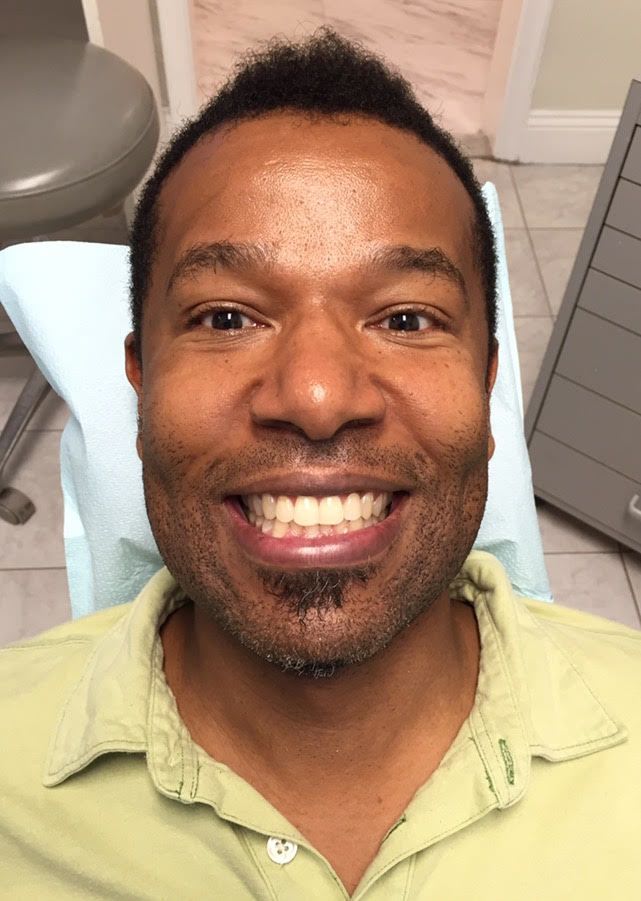
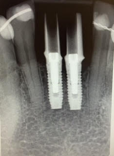
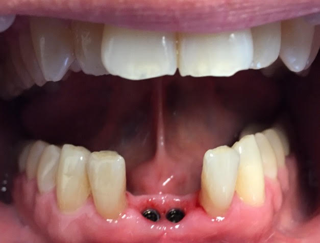
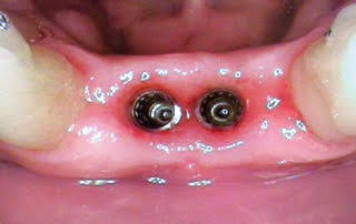
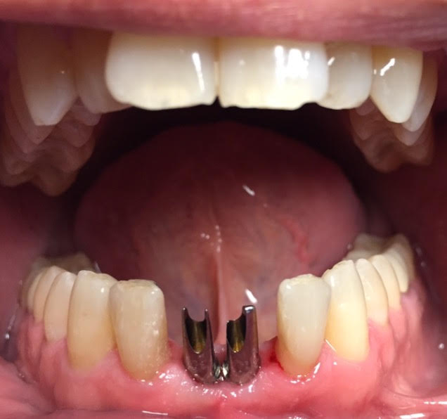
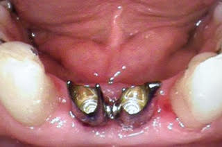
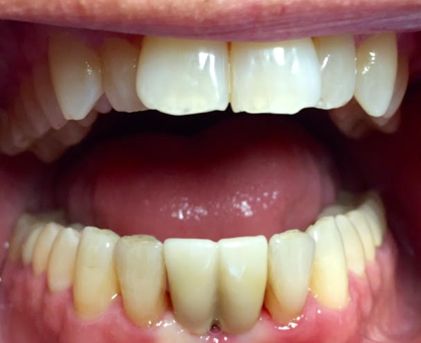
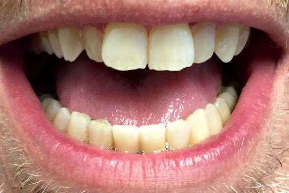
This patient lives in Ft. Lauderdale and flew up today for me to restore his lower front teeth with dental implant crowns. This was a challenging case because of how thin the bone was and the tight space, and close proximity of the implants.
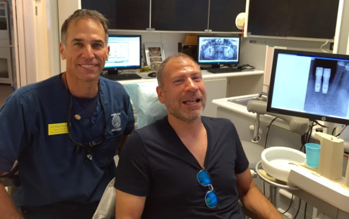
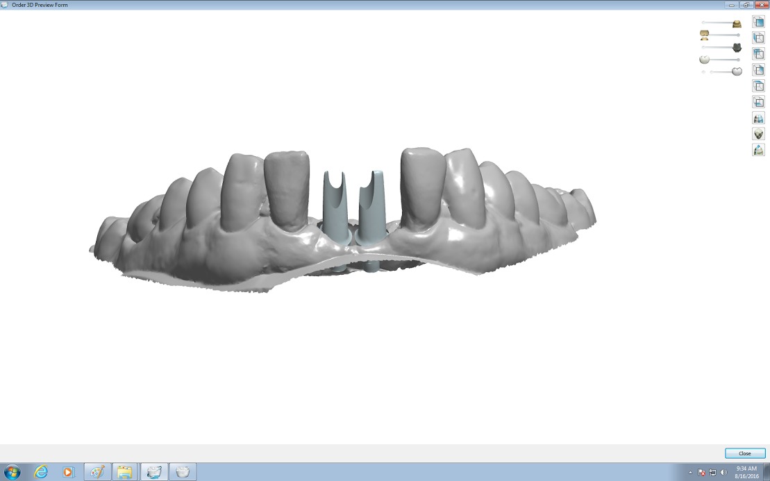
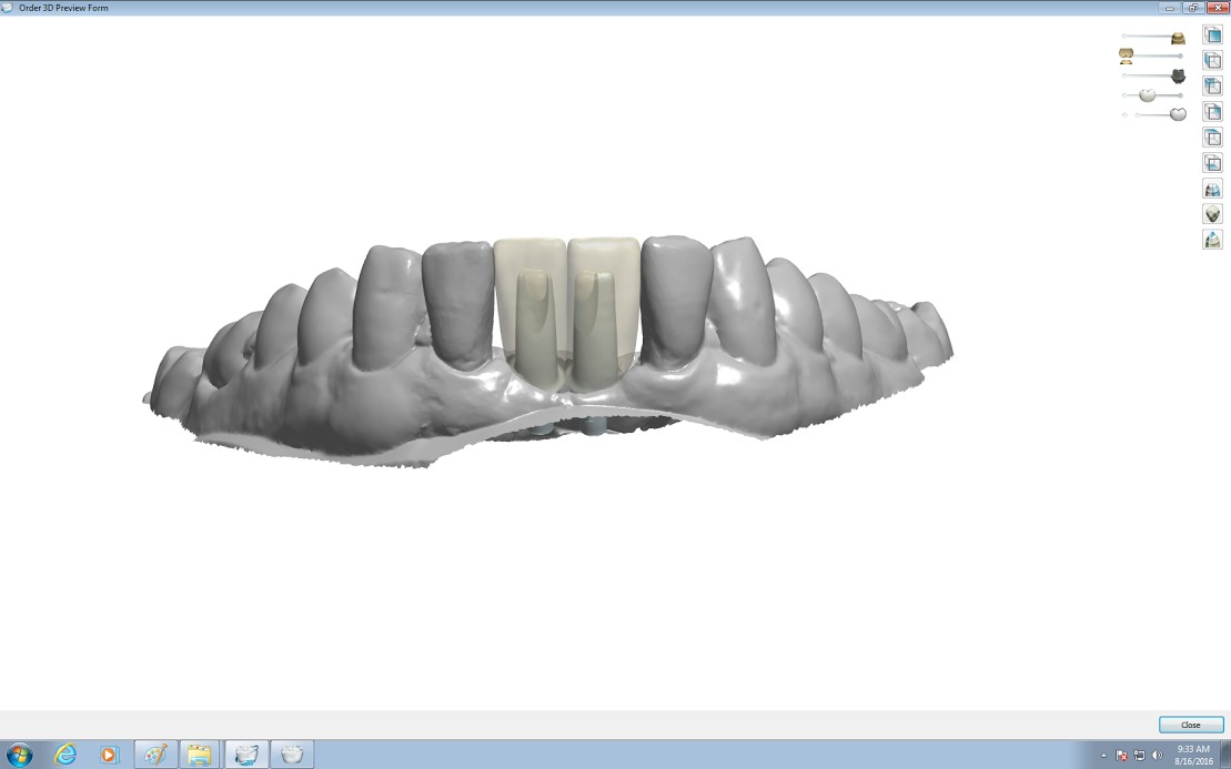
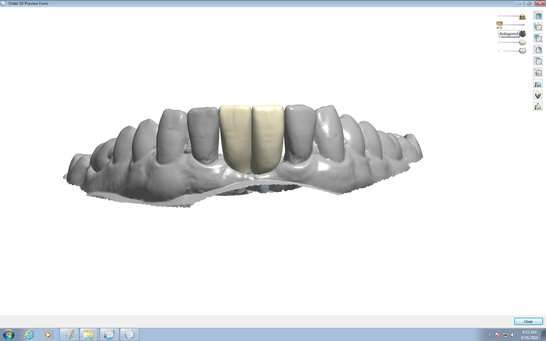
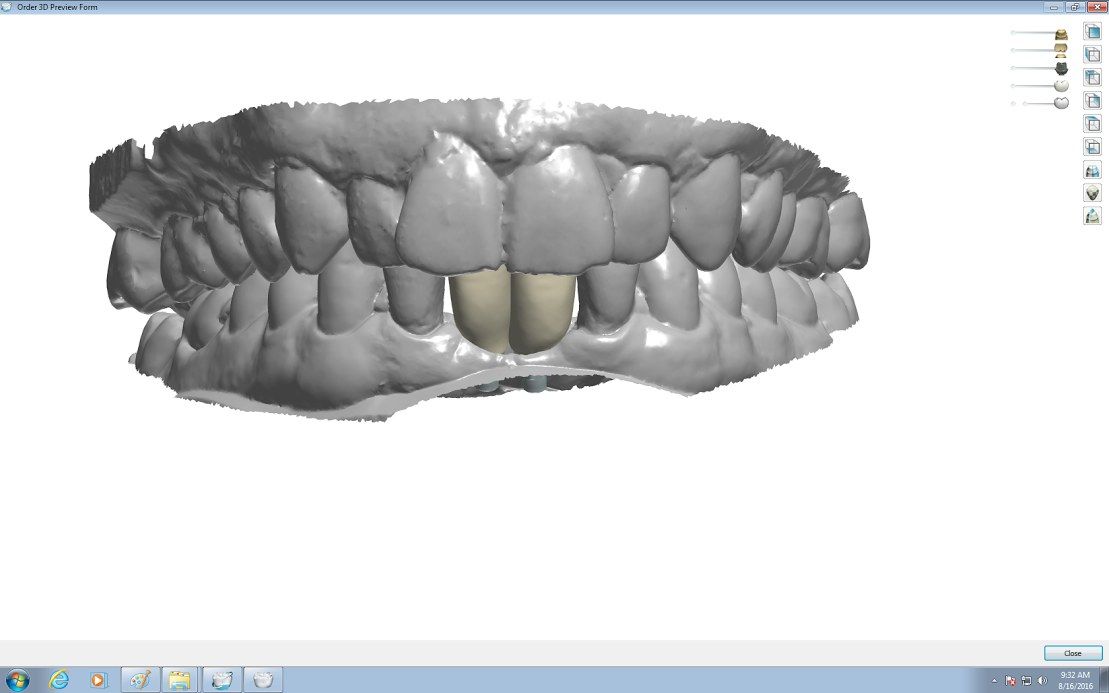
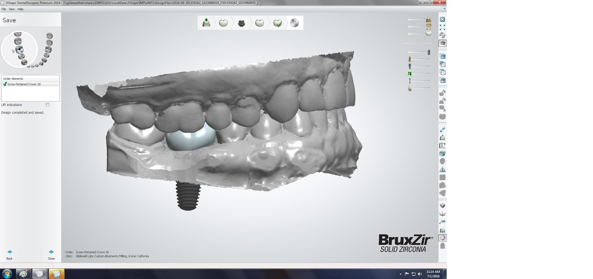
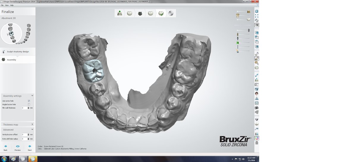
digital design scans
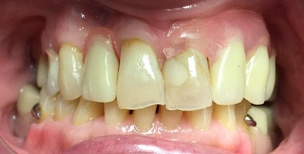
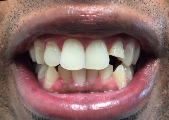
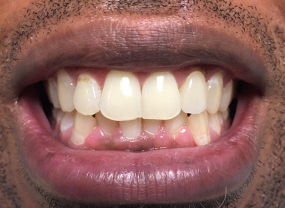
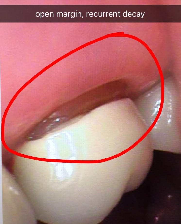
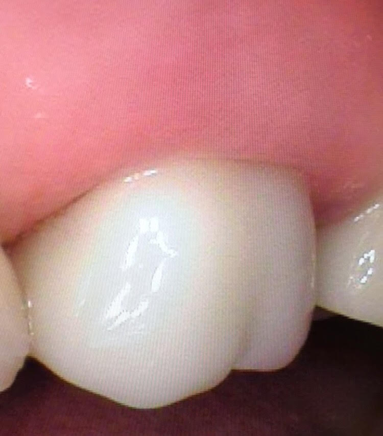
This patient wanted me to replace her 20 year old crown. She had gum recession exposing her root and recurrent decay that had spread under her old crown. The dark area showed when she smiled. I cut off the old crown, removed the decay and made her a beautiful new porcelain crown to cover the exposed root. Here’s the before and after photos.
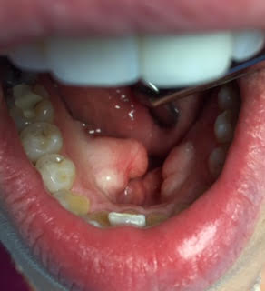
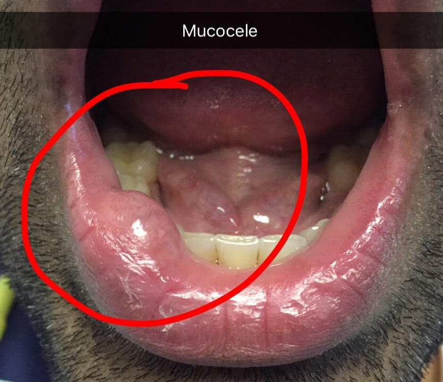
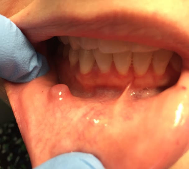
These are 2 patients have swellings on the inner surface of the lower lip and cheek. The first is a 31 year old male, that has had it for 1 month. The second is a 27 year old female that has had it for 2 weeks. This is called a mucocele. It is caused when you bite or traumatize your lip and rupture or obstruct the small salivary gland duct. The saliva pools up and causes a small swelling. There are many small salivary glands on the inner surface of the lip and damaging the duct can cause the saliva to pool up causing a clear or bluish swelling. My treatment for this condition is to have the patient rinse with warm salt water 6 times a day for 1 week. If it is still present after 1 month have it surgically removed.
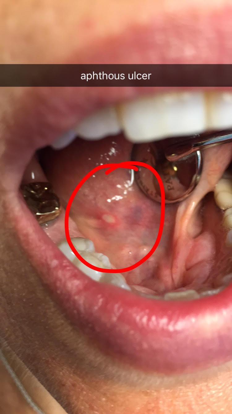
This next condition on this 55 year old female patient is called an aphthous ulcer, sometimes referred to as a canker sore. This is usually caused by stress, not eating or sleeping enough and getting run down. I see this a lot in college kids who are stressed during finals and not sleeping enough or eating well. It can also be caused by an allergic reaction. The aphthous ulcer usually resolves on it’s own in 2 weeks and can reoccur. It can sometimes be painful, and if it’s really bothering the patient, I can prescribe a Chlorohexidine mouthwash or steroid cream.
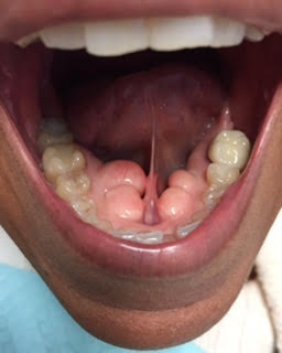
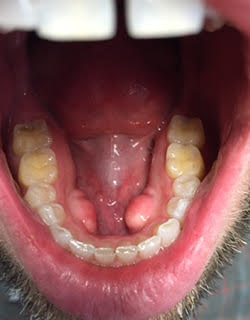
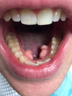
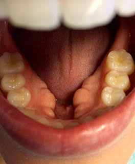
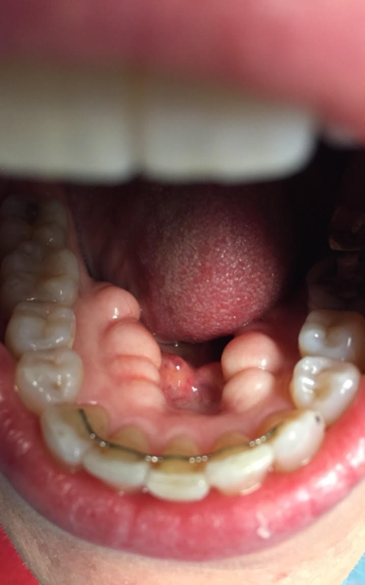
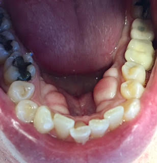
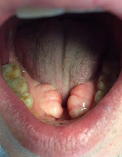
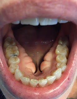
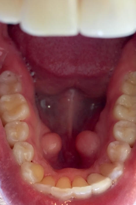
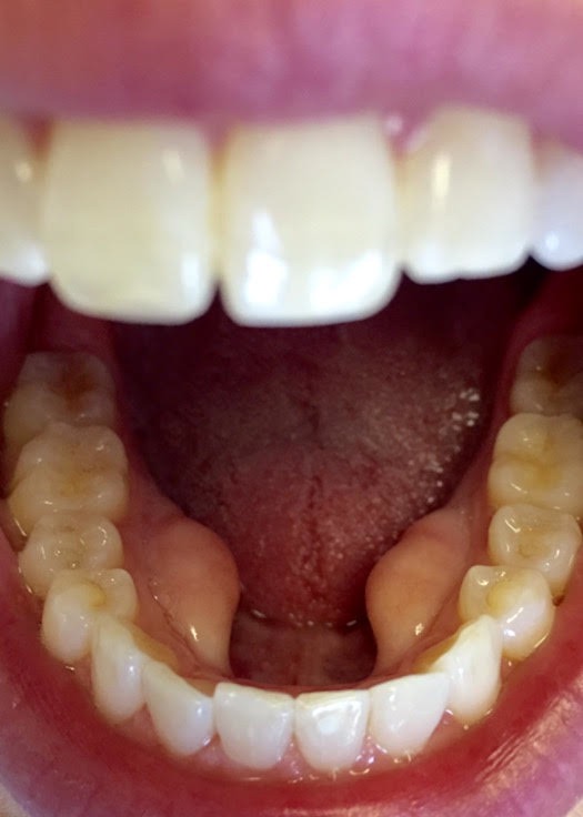
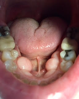
These next patients have mandibular tori, also called torus mandibularis. A torus (plural tori) is a bony growth that occurs in the lower or upper jaw in 20% of the population. Tori are normal bone covered by normal tissue. These are usually caused by genetic factors and teeth grinding causing excess bone to be deposited. No treatment is required.
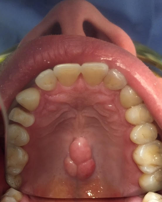
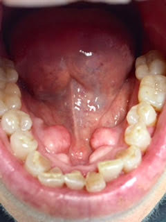
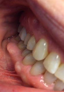
This last patient has both maxillary and mandibular tori: 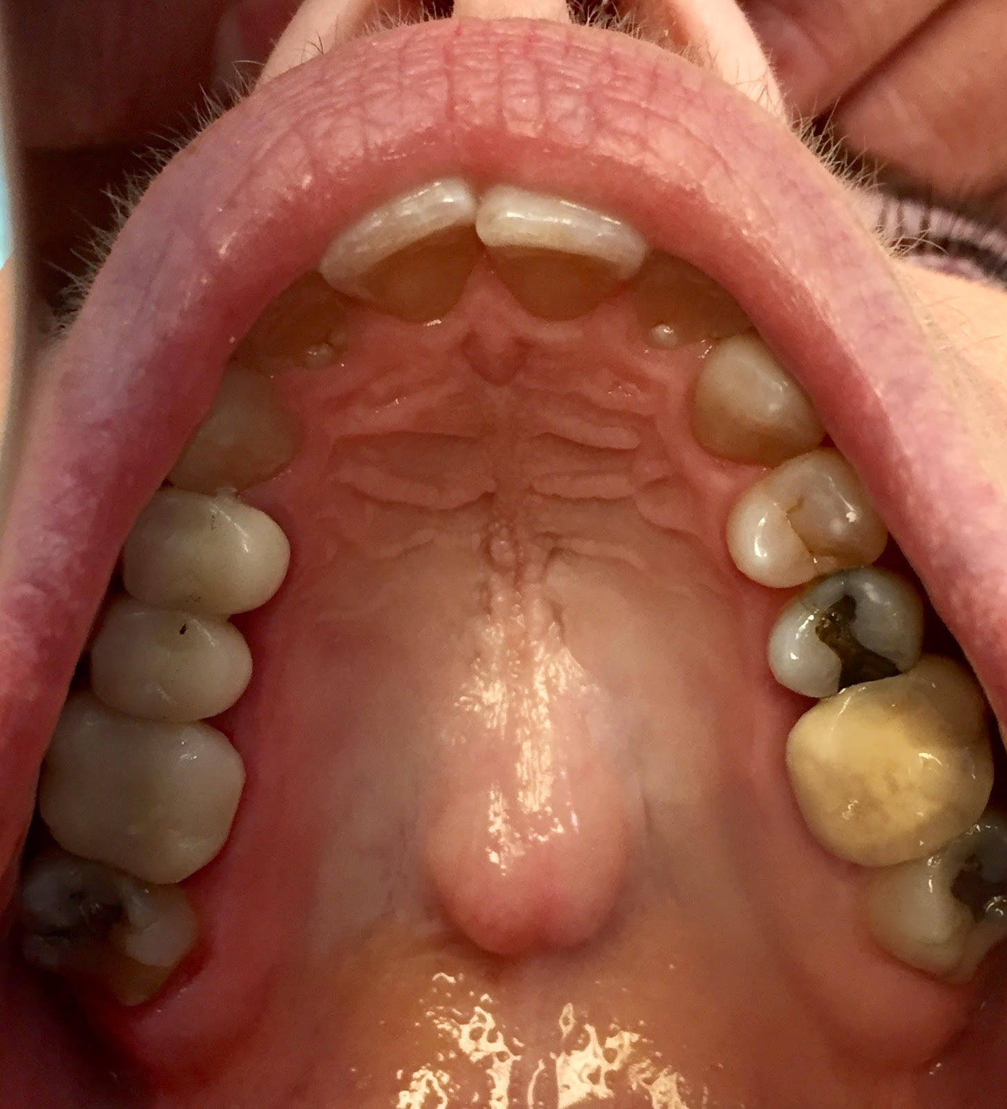
This patient’s upper left first molar tooth was fractured and very painful. We extracted her tooth and placed a Biomet Zimmer Encode Implant. Today I finished restoring her molar with a beautiful dental implant.
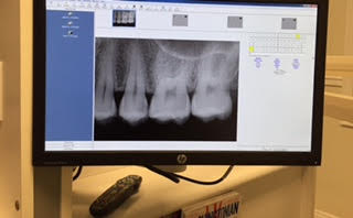
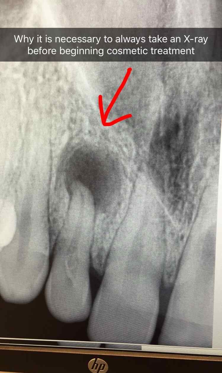
This 19 year old was home from college for a few weeks and wanted me to do veneers to fill in the spaces between her upper front teeth and give her a pretty smile. I took x-rays prior to starting treatment and noticed a large abscess at the root apex of this lateral incisor tooth. The tooth was asymptomatic and the patient said the tooth never hurt or felt sensitive. The tooth must has been traumatically injured when she was young. It’s good we discovered this abscess and can take care of it before it destroys more bone and adjacent teeth.
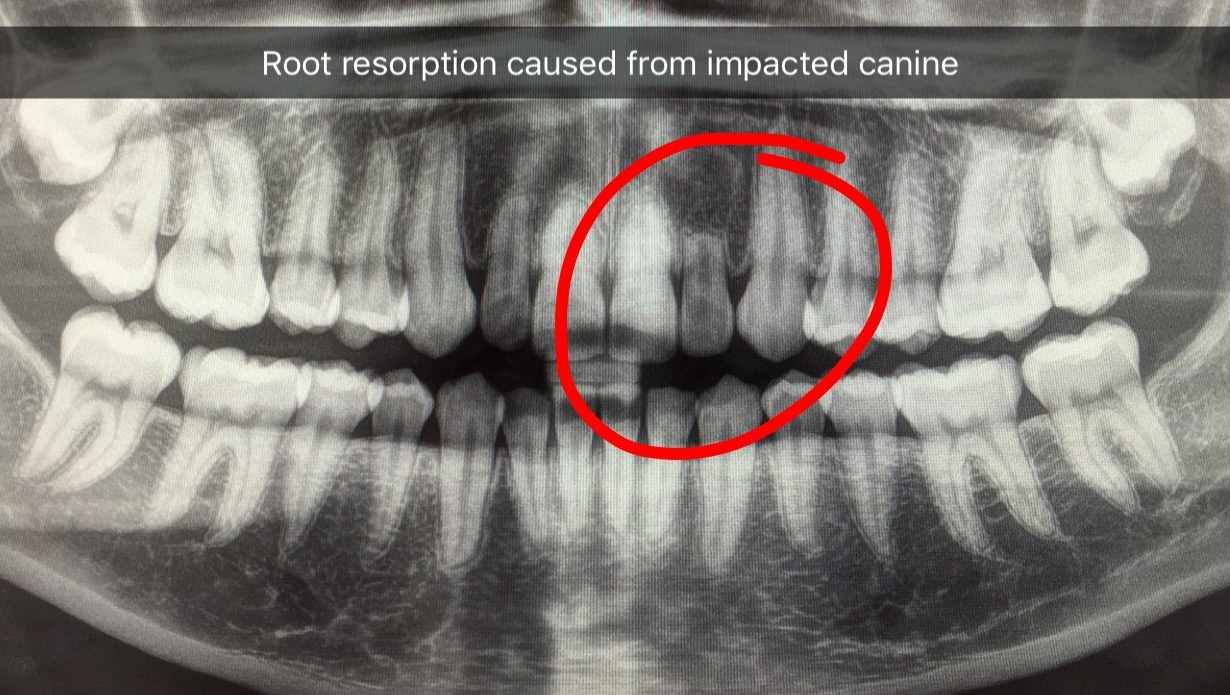
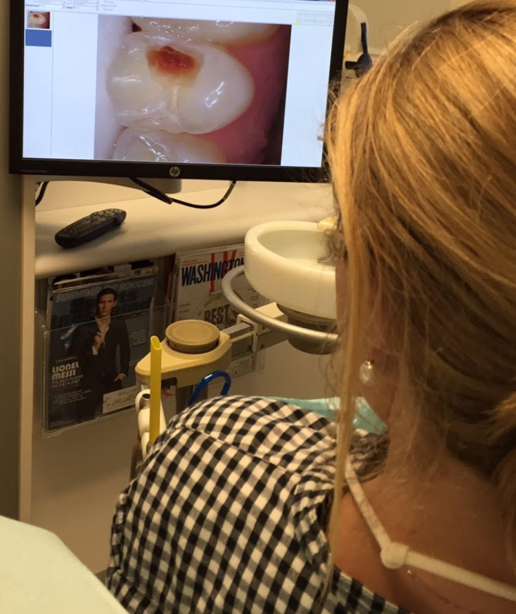
At the same time in a different dental operatory was this other patient who was here for her cleaning and check-up appointment. Notice the resorbed root on the maxillary lateral incisor. The root is about half as long as it should be. This was caused by her impacted canine tooth, stuck above this root, that was later brought down into place orthodontically.