Importance of taking a radiograph to verify accurate interface of the custom abutment to implant.

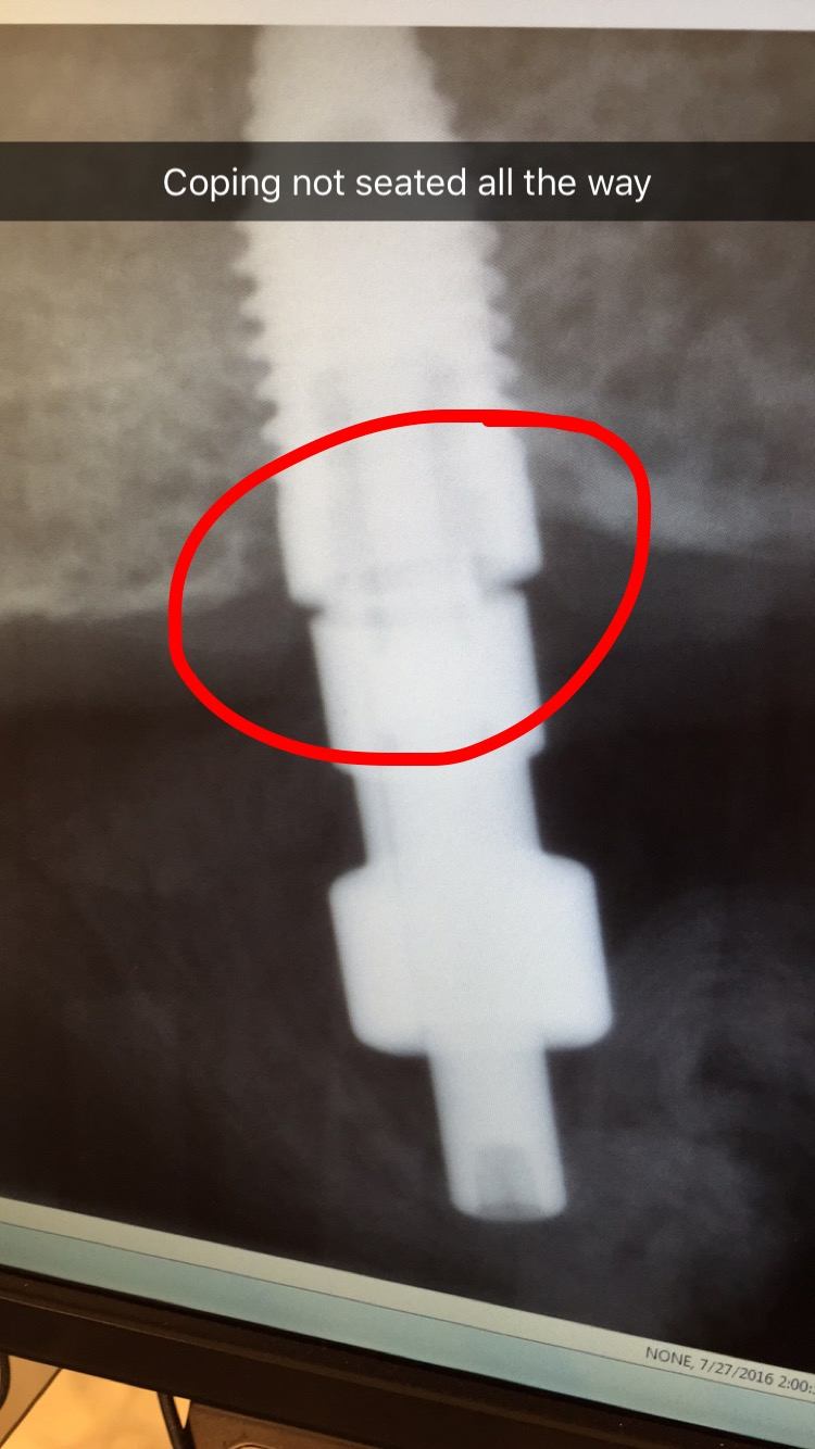
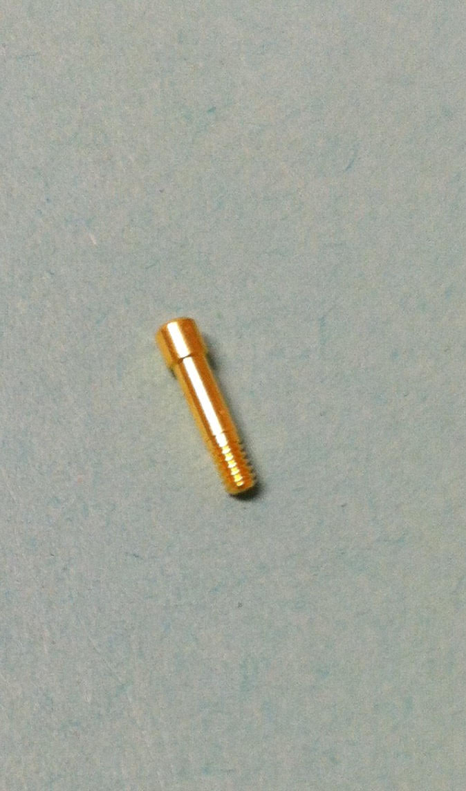
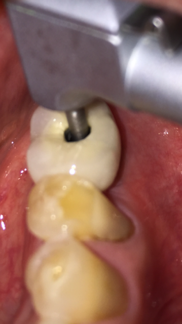
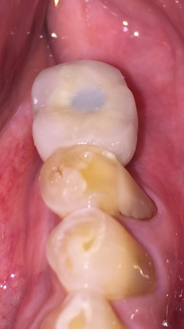
In this case report Dr. Gentry demonstrates the importance of taking a radiograph when seating the custom abutment and crown before final torquing of the gold implant interface connecting screw.

unnamed
Implant
unnamed-1
Custom abutment and crown placed with gold screw tightened to finger pressure.
unnamed-9
Upper radiograph shows improper interface-note the gap present at the interface. Lower radiograph shows perfect fit, taken after adjusting the contact and re-positioning the crown/abutment on the implant.
unnamed-10
Gold screw used to attach crown and abutment to implant.
unnamed-2
Now the gold screw can be tightened to 20 Newtons per centimeter squared with the torque wrench.
unnamed-3
Screw hole filled in with composite filling material.

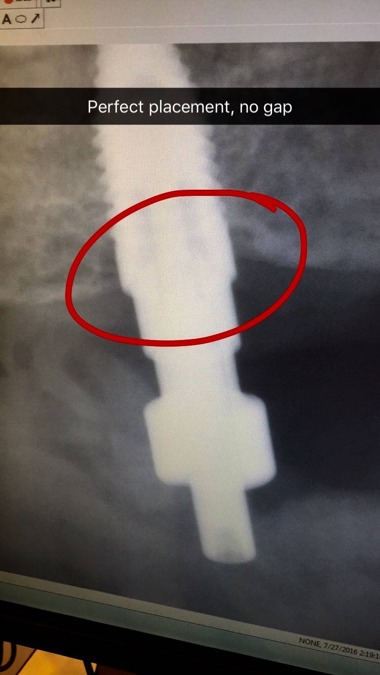
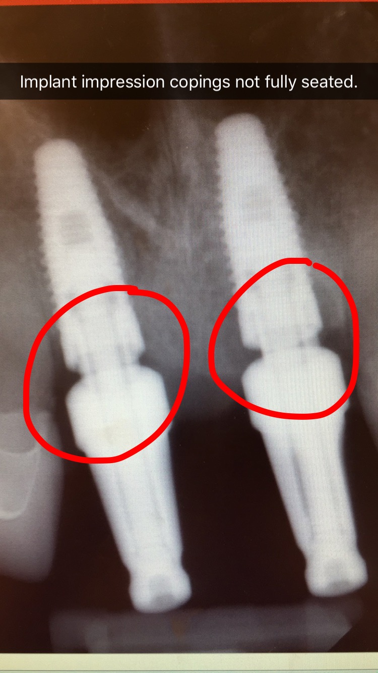
Here’s an example from a case I was doing with a dental student involving the 2 upper front teeth. In the first x-ray the impression copings were not fully seated. I re-positioned the copings and another x-ray was taken to confirm proper alignment. You cannot tell without an x-ray since this is below the gum-line.

implantseating1
x-ray verification showing implant copings not fully seated
implantseating2
After re-positioning, perfect seating

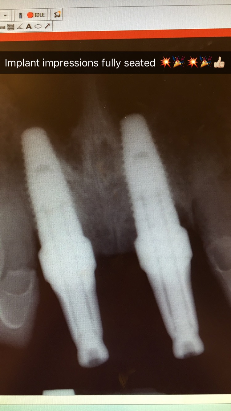
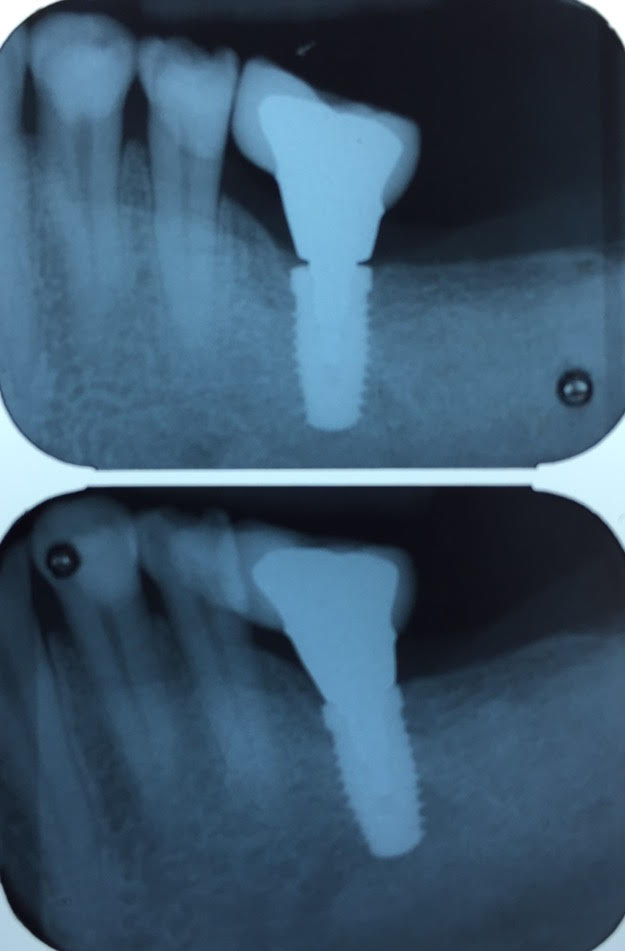
Here’s an example of another implant check x-ray to verify proper seating.

coping xraycoping xray2